High NA Optical Fiber Microscope
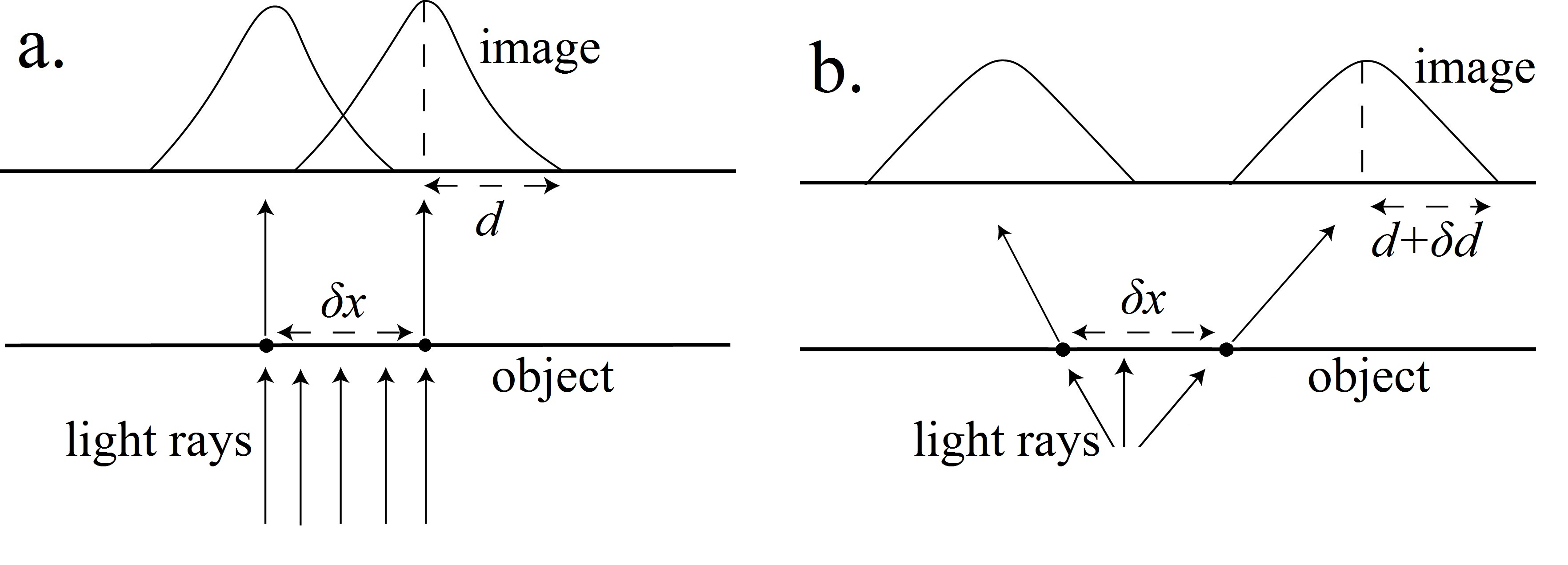
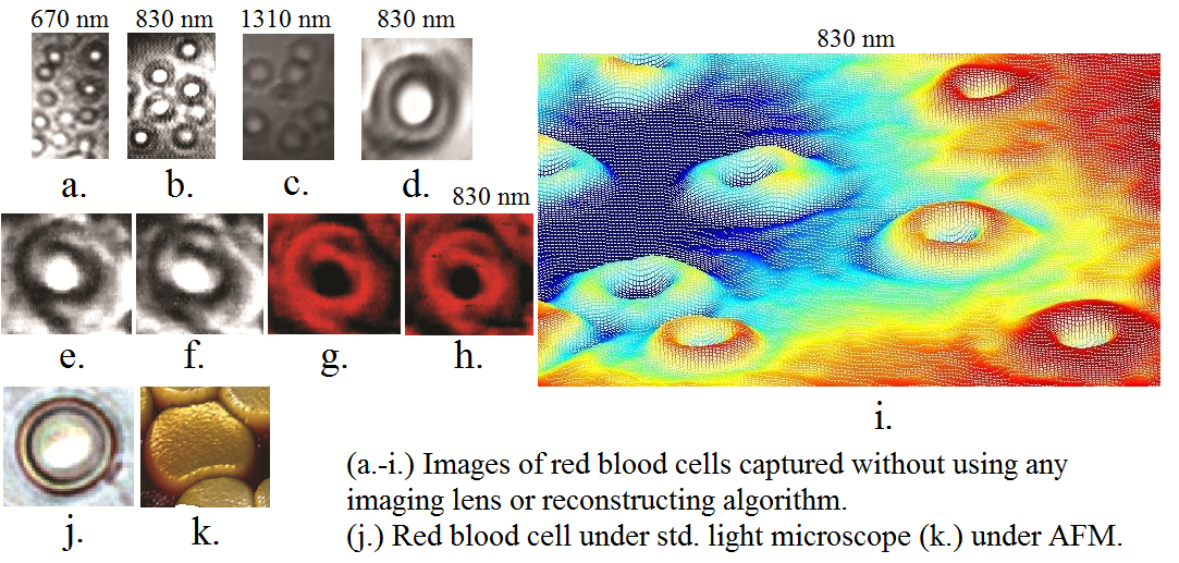
We proposed a non-computational method of counteracting the effect of image degradation introduced by the diffraction phenomenon in lensless microscopy. All the optical images (whether focused by lenses or not) are diffraction patterns, which preserve the visual information upto a certain extent determined by the size of the point spread functions, like airy disks in some cases. A highly diverging beam can be exploited to reduce the spatial extent of these point spread functions relatively in the transformed projective space, which can help us in the spatial unmixing of the visual information. The principle has been experimentally validated by the lensless imaging of red blood cells of diameter ~6-9 micrometers and a photolithography mask with features in micrometer scale. These lensless images were captured using imaging pixels of size ~18 micrometers i.e. pixels were several times larger than the target object(s) itself. The important advantages of the proposed approach of non-computational shadow microscopy are the improved depth of field and a drastic increase in the sensor to sample working distance. The imaging method can also be used as a projection technique in the multi-angle optical computed tomography (CT). For more details and results check this>> Link and Link.

